Ding’s group developed 3D printing of biomacromolecular porous scaffolds using “nascent hydrogel” of modified gelatin for osteochondral regeneration
The cartilage is, once damaged, hard to self-heal due to the lack of blood supply, etc., and it is more challenging to repair an osteochondral defect concerning both cartilage and subchondral bone. Herein, it is hypothesized that a bilayered porous scaffold composed of a biomimetic gelatin hydrogel might, despite no external seeding cells, induce osteochondral regeneration in vivo after being implanted into mammal joints.
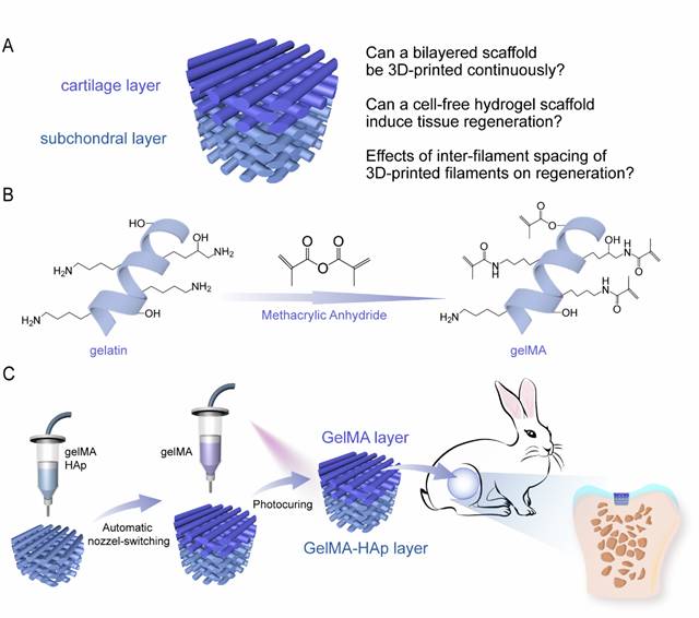
Figure 1. Schematic diagram of the bilayered scaffold and the one-step 3D-printing of a bilayered hydrogel scaffold for osteochondral regeneration.
This idea was confirmed based on the successful continuous 3D-printing of the bilayered scaffolds combined with the sol-gel transition of the aqueous solution of a gelatin derivative (physical gelation) and photocrosslinking of the gelMA (gelatin methacryloyl) macromonomers (chemical gelation). At the direct printing step, a nascent physical hydrogel was extruded, taking advantage of non-Newtonian and thermoresponsive rheological properties of this 3D-printing ink. In particular, a series of GelMA (crosslinked gelMA) and GelMA-HAp (hydroxyapatite) bilayered hydrogel scaffolds were fabricated to evaluate the influence of the spacing of 3D-printed filaments on osteochondral regeneration in a rabbit model. The moderately spaced scaffolds outputted excellent regeneration of cartilage with cartilaginous lacunae and formation of subchondral bone. Thus, tricky rheological behaviors of soft matter could be employed to improve 3D-printing, and the bilayered hybrid scaffold resulting from the continuous 3D-printing is promising as a biomaterial to regenerate osteochondral articular cartilage.
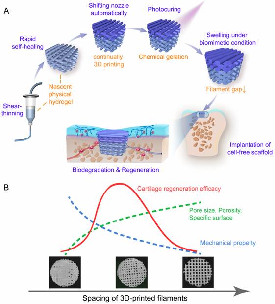
Figure 2. Schematic diagrams of the key material properties to enable the continuous 3D printing of the present bilayered hydrogel scaffold and the cues underlying the effects of inter-filament spacing on osteochondral and particularly cartilage regeneration.

Figure 3. Optical micrograph of HE-stained slice in the site of regenerated cartilage in femoral trochlear of rabbits at 12th week using the optimal bilayered scaffold. This image is a mosaic of 15 photographs (5 times 3).
This work was carried out via cooperation between Ding’s group from Fudan University and Chen’s group from Huashan Hospital. The results have been published on Advanced Healthcare Materials with a PhD candidate student Jingming Gao and Dr. Xiaoquan Ding as the first authors. See please: Jingming Gao#, Xiaoquan Ding#, Xiaoye Yu, Xiaobin Chen, Xingyu Zhang, Shuquan Cui, Jiayue, Shi, Jun Chen, Lin Yu, Shiyi Chen*, Jiandong Ding*, Cell-free bilayered porous scaffolds for osteochondral regeneration fabricated by continuous 3d-printing using nascent physical hydrogel as ink, Advanced Healthcare Materials, 2020: e2001404
Article links: https://doi.org/10.1002/adhm.202001404