“Critical” Areas of Cell Adhesion on Micropatterned Surfaces
The adhesive area is important to modulate cell behaviors on a substrate. A corresponding methodology paper was published as follows:
Ce Yan, Jianguo Sun, and Jiandong Ding*, Critical areas of cell adhesion on micropatterned surfaces, Biomaterials, 32, 3931-3938 (2011)

We spent 6 years to make this study. In contrast, it took just 5 days from manuscript submitting to acceptance for publication.
This paper aims to semiquantitatively examine the existence of the characteristic areas of cell adhesion on the level of individual cells. We prepared a series of micropatterned surfaces with adhesive microislands of various sizes on an adhesion-resistant background, and cultured cells of MC3T3-E1 (osteoblast), BMSC (bone mesenchymal stem cell) or NIH3T3 (fibroblast) on those modeled surfaces.
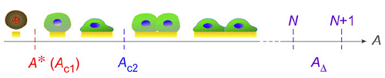
We have defined seven characteristic areas of an adhesive microisland and confirmed that they are meaningful to describe cell adhesion behaviors:
(1) the critical adhesion area from apoptosis to survival denoted as A* or Ac1;
(2) the critical area from adhesion of a single cell to adhesion of multiple cells (Ac2);
(3) the basic area for one more cell to adhere (AD);
(4) and (5) the characteristic areas of a microisland most probably occupied by one cell (Apeak(1)) and two cells (Apeak(2));
(6) and (7) the characteristic areas of a microisland occupied by one cell (AN(1)) or two cells (AN(2)) on average.
We further discussed the relationship between those characteristic areas and the spreading area on a non-patterned adhesive surface.